

“No great discovery was ever made without a bold guess.”
― Isaac Newton


Our current discovery research projects are centered on understanding light-matter interaction for sensing and exploring biological processes.
Problem
Developing societies face significant challenges in accurately detecting and effectively treating diseases, largely due to limited access to diagnostics.
This issue is compounded by the overuse of broad-spectrum antibiotics, often administered without proper diagnostic procedures.
To address this, a comprehensive understanding of diseases and pathogens, their metabolic processes, and relevant biomarkers is essential for
creating affordable and easy-to-use diagnostic and treatment methods.
Many biomolecules, upon absorbing light, become excited and then rapidly relax, re-emitting light at a lower energy. This emitted light, called fluorescence, offers crucial insights into the molecules. While reagents are often used to boost fluorescence for studying minute molecular quantities, our research focuses on developing reagent-free methods. We aim to achieve this through several approaches, including: (a) improving instrumentation, and (b) gaining a deeper understanding of energy exchange between biomolecules, allowing us to probe the same biomolecules through various interactions with different molecules.
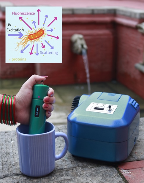
Fluorometric systems with sub-ppb sensitivity,
developed using our groundbreaking methods.
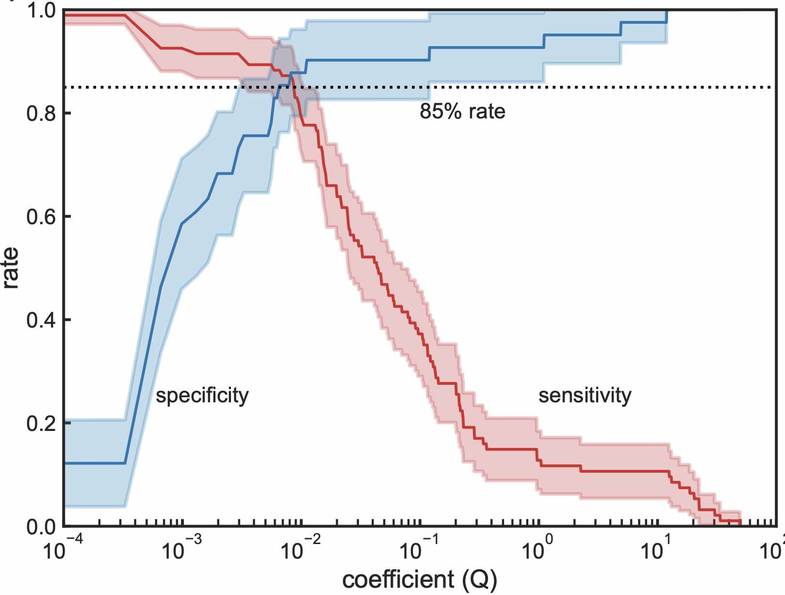
Our new analytical method identifies fecal coliforms with unprecedented
sensitivity and specificity.
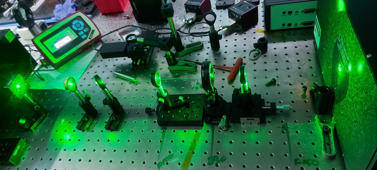
Raman spectroscopy offers a non-distructive, reagent-free, specific, and broadly applicable method for examining the molecular characteristics of both living and non-living matter. Our research aims to apply this technique, combined with optical trapping methods, to investigate live bacterial and virus particles. We use two optical trapping methods:
While optical trapping within a micro-Raman setup is now a relatively standard procedure, a
fundamental physical limitation known as the diffraction limit has historically resulted in Raman signals being a
composite of signals originating from all subcellular structures within the measurement volume and their surroundings.
Consequently, achieving high specificity and extracting information from the subcellular structures of live bacterial
cells via Raman spectroscopy has remained a significant challenge.
Our theoretical investigations into Brownian motion within an optical trap have revealed that different subcellular
structures, possessing distinct optical and hydrodynamic properties, exhibit varying Brownian motion patterns within the cells.
This differential motion leads to varying average Raman signal amplitudes when the excitation light is modulated. We intend to
leverage these differences in signal amplitude to pinpoint and analyze the various subcellular structures of a live bacterial
cell captured within an optical field.
Our research utilizes the evanescent optical field near photonic waveguides to form an optical trap for Raman spectroscopy.
This technique generates an attractive gradient force, enabling the manipulation of microorganisms and viruses in the transverse direction,
while a propulsive scattering force facilitates their movement along the longitudinal path of light propagation.
These fully integrated Raman sensing circuits offer significant advantages. They can be mass-produced for a few dollars,
providing a low-cost solution. Furthermore, these systems are capable of amplifying the Raman signal by up to four orders of magnitude.
If successful, this technology holds the potential to revolutionize diagnostics in resource-limited areas, particularly within developing countries.

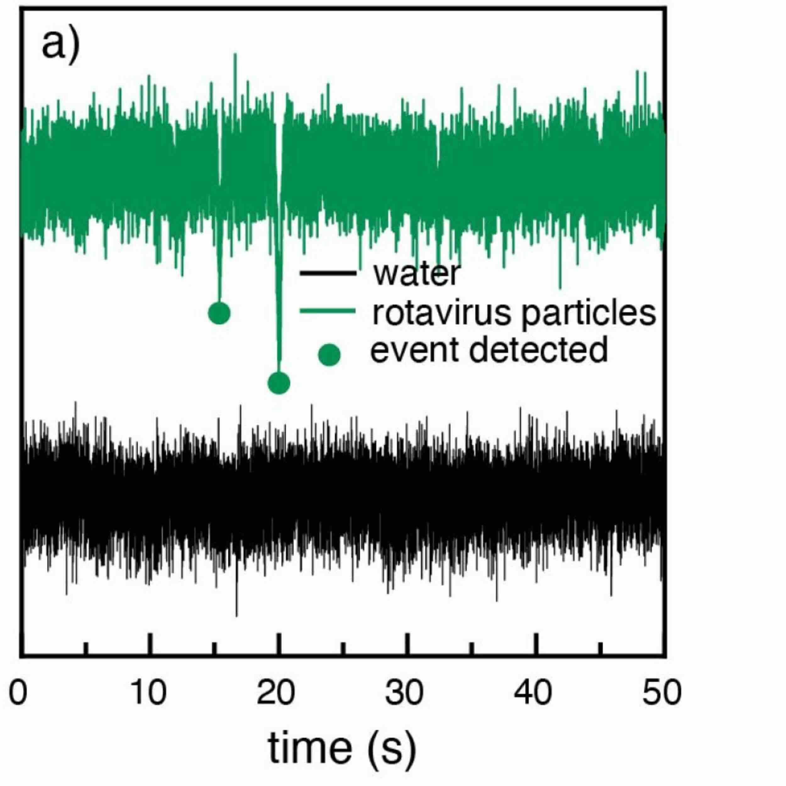
Signals from rotavirus particles measured using light scattering.
Studying microorganisms typically requires expensive and complex microscopy systems, which are not easily accessible in developing countries. However, we've found that it's possible to gain specific information about these organisms without necessarily imaging them in their real-life forms. Our approach utilizes Diffuse Correlation Spectroscopy (DCS), a computational optics technique. DCS works by sensing minute changes in the path of a light beam through laser speckle analysis. This method has been successfully applied by our partners at the University of Glasgow for heart and brain diagnostics. We are now adapting DCS to detect and study microorganisms, particularly those responsible for diarrheal diseases.
Problem:
Photonics circuits have been traditionally developed using nano-lithographic techniques, which either use
lithographic masks or e-beam writing followed by dry-etching methods. Both of these approaches are expensive to be
implemented in resource-limited settings, which therefore has hindered photonic innovations in developing countries.
We are exploring methods to develop a wet-etching method assisted by laser pulses.
Femtosecond Laser Writing (FLW) creates photonic structures by focusing a high-energy femtosecond laser beam to alter a material's refractive index. While FLW is demonstrated, the underlying physical processes are poorly understood. This project aims to develop a physical model for glass densification induced by femtosecond laser radiation, using pump-probe Raman and Brillouin spectroscopy. This model will be crucial for creating reproducible, high-quality photonic components on glass, benefiting photonics applications, especially in developing countries.
Problem:
Animals possess an astonishing ability to detect extremely low concentrations of chemicals,
with a response time and sensitivity that far surpass any human-made tools. Imagine a device capable of "smelling" diseases
like cancer or diabetes by identifying relevant chemicals in urine or breath, much like trained dogs. Such technology could
revolutionize diagnostics in developing nations.
A highly debated but promising theory suggests that olfaction (smelling) occurs when the vibrational energy of an odorant aligns with the electronic transitions of olfactory receptors, enabling quantum tunneling of electrons through the odorant. Regardless of whether this process is truly involved in natural olfaction, its principles could be applied to develop highly sensitive chemical detection methods. Our research focuses on building a pump-probe based Ultrafast Raman spectroscopy setup. This will allow us to investigate, with femtosecond resolution, the temporal dynamics of interactions between the vibrational energy of an odorant and tunable "artificial receptors."